Unraveling the Aging Puzzle: AI and a Curious Mind Connect the Dots
Jeff T. Bowles and Grok 3.0 3/5/2025
Key Points
- Research suggests a rise in LH, hCG, and possibly FSH likely leads to increased short LARP1, driving aging processes.
- It seems likely that FSH triggers aging symptoms in systems #1 and #2, more associated with general body aging, while LH and hCG trigger systems #3 and #4, linked to reproductive and genetic stability aging.
- The evidence leans toward hormonal changes, particularly during and after puberty, influencing short LARP1 and aging, with SP-1 playing a key role in linking sex and aging.
Hormonal Triggers and Aging Systems
Aging involves complex hormonal shifts, and it appears that LH, hCG, and possibly FSH all contribute to increasing short LARP1, a molecule that coordinates four aging systems. FSH seems to be more involved in systems #1 and #2, which affect vascular and mitochondrial functions, often seen as more “male-like” aging patterns. On the other hand, LH and hCG are likely more linked to systems #3 and #4, involving DNA repair and reproductive aging, potentially more “female-like.” This distinction helps explain how aging differs between genders, though both experience these changes.
The Role of SP-1
An unexpected detail is that SP-1, one of Horvath’s aging genes, not only regulates death genes MAO-A and MAO-B but also triggers WRN production during puberty. This connects sexual maturation to aging, showing how puberty sets the stage for later life decline, tying sex and aging into a single system.
Connecting the Dots
Research suggests these hormonal increases, especially after age 50, create a pro-inflammatory, free radicalized, acidic environment, boosting short LARP1 and driving aging. This process starts with puberty, where surges in FSH, hCG and LH surges apparently activates SP-1 which in turn activates WRN, and continues with hormonal surges of testosterone and estradiol amongst other hormones impacted, affecting how our cells develop and age over time.
Survey Note: Integrating Hormonal Changes and Short LARP1 into the Unified Theory of Aging
This analysis integrates four blog posts and additional user-provided information to propose a comprehensive theory of aging, emphasizing how a rise in luteinizing hormone (LH), human chorionic gonadotropin (hCG), and possibly follicle-stimulating hormone (FSH) initiates increased expression of short LARP1, and how these metabolic changes fit into the evolutionary, metabolic, epigenetic, developmental ,and molecular dimensions of aging. The posts collectively suggest that aging is a programmed, multi-layered process, with specific molecular mechanisms driving its progression. Below, we detail the synthesis, drawing on the insights from each post and the user’s additional facts, identifying hidden relationships and focusing on the initiation of GABA and αKG decline, with a focus on hormonal influences and SP-1’s role.
Background and Context
The first blog post, “AI’s Deep Analysis of Horvath’s 48 Aging-Related Genes Across Biological Processes” Horvath’s Epigenetic Clock Analysis Across Biological Processes, explores Stephen Horvath’s epigenetic clock, which identifies 48 genes whose DNA methylation changes correlate with aging across mammals. These genes, including transcription factors like REST and NANOG, and splicing regulators like CELF4, are implicated in metabolism, stress response, mRNA splicing, and development. The post highlights the interplay between metabolic changes (e.g., declining α-ketoglutarate, αKG) and epigenetic modifications, noting enzymes like monoamine oxidase-A & B (MAO-A & B) and CD38 that become dysregulated with age, depleting essential cofactors like NAD⁺ and FAD, and exacerbating cellular aging.
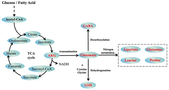
The second post, “Metabolic Imbalance of the GABA–Glutamate–αKG Axis in Aging” Metabolic Imbalance and Aging GABA–Glutamate–αKG Axis, delves into how aging shifts neurotransmitter, serum, and metabolic profiles, with serum γ-aminobutyric acid (GABA) declining, glutamate increasing, and αKG levels dropping. These changes disrupt neurotransmitter and serum balance, impair mitochondrial function, and affect epigenetic regulation through inhibition of αKG-dependent demethylation, linking metabolism to Horvath’s epigenetic clock.
The third post, “The Four Horsemen of Aging: How 4 Evolved Mammalian Aging Systems Reveal the Missing Half of Evolution” Four Horsemen of Aging Evolutionary Systems, proposes that aging comprises four independently evolved systems, each co-opting the vulnerabilities of the last. These systems are tied to evolutionary stages: System #1 (plant-like vascular decline), System #2 (mitochondrial dysfunction in motile animals), System #3 (DNA repair and immune function), and System #4 (sexual reproduction, exemplified by Werner’s syndrome which is triggered by puberty). It introduces short LARP1, a nuclear lncRNA, as a putative potential orchestrator, mis-splicing or suppressing mRNAs of protective proteins like WRN, ATM, and XP/CS.
The additional post, “MAO-B Forces a Rethink of Darwin’s Legacy” MAO-B Forces a Rethink of Darwin’s Legacy, discusses Monoamine Oxidases A and B (MAO-A and MAO-B) as potential “death genes” that increase with age and deplete FAD, mirroring the way CD38 depletes NAD⁺. It challenges traditional evolutionary theory by suggesting that MAO-B is a true death gene with no early-life benefit, proposing a programmed aging model.
The user provided additional information, including:
- Both GABA and αKG declines are involved in increasing M1 macrophages or their production of CD38, with αKG decline shutting down about 36 of Horvath’s 48 genes (noted in his first draft of his seminal paper-the identified gens have changed somewhat in later revisions).
- Serum αKG declines by about 90% or more with aging, and serum GABA declines, though the extent is unclear.
- After age 50, LH, FSH, and hCG increase by hundreds to thousands of percent, with FSH increasing more in men and LH more in women. With aging, LH molecules become larger, have a longer half-life, and may become highly acidic, potentially also applying to FSH. Interestingly large surges in these hormones occur during fetal development, and initiation of puberty, suggesting that aging and development appear to be part of the same programmed process.
- LH and hCG are associated with cancers and assigned to aging systems 3 and 4, while FSH is not associated with cancers and assigned to systems 1 and 2. These hormones may increase free radicals and isoforms become larger, more acidic, more bioactive and immunoactive, and have longer half lives with aging.
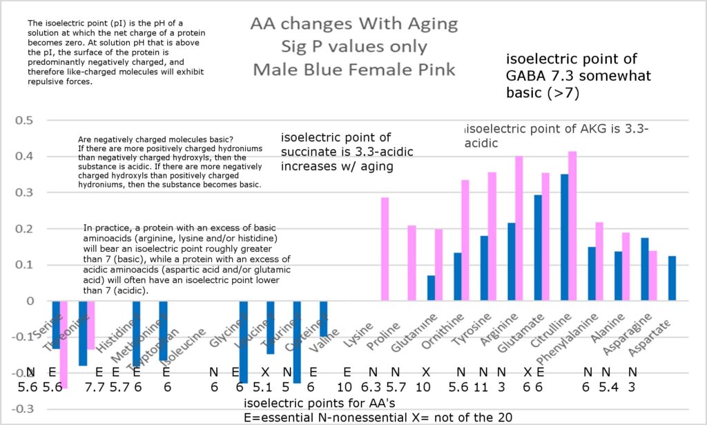
- Melatonin, dehydroepiandrosterone (DHEA), and pregnenolone, with antioxidant properties, decline dramatically with aging (up to 90%), and progesterone drops to almost 0 in menopausal women, later in men. These antioxidant hormones might be basic (high pH), while pro-aging hormones are associated with cAMP, and antioxidants with cGMP stimulation.
- Elevated LH is associated with Alzheimer’s, frailty, tissue atrophy, and cancer, while elevated FSH is linked to abdominal obesity and heart disease.
- The user hypothesizes that increasing hormonal acidity affects intracellular pH, lowering it and reducing GABA and αKG production and increasing glutamate by altering enzyme activity, .
- Of Horvath’s 48 genes, 12 lose methylation at a single locus in Horvath’s first draft, including LARP1, some related to Alzheimer’s and circadian rhythms (which suggests a melatonin connection) , LHFLP3, LHFLP4, and others. Antioxidants catalyze methylation of cytosine normally, (except in cases where the reverse acting acting TDG and TET enzymes are involved), while free radicals normally catalyze demethylation of 5mC.
- The user requested to attribute the triggering of short LARP1 to LH, hCG, and possibly FSH, with FSH more related to aging systems #1 and #2 (male-like aging) and LH and hCG to systems #3 and #4 (female-like aging).
- SP-1, one of Horvath’s additional aging genes uncovered in the third revision of his seminal paper, binds to the promoters of MAO-A and MAO-B, and also triggers the production of WRN in various tissues for the first time when puberty hits, tying sex and aging into one highly related system.
Synthesis: A Unified Theory
Our unifying theory posits that aging is a programmed process with four evolutionary layers, interconnected through metabolic and epigenetic mechanisms, with short LARP1 serving as a central regulator, and death genes like MAO-A, MAO-B, and CD38 exacerbating the process through cofactor depletion. The decline of GABA and αKG is initiated by these systems and hormonal changes, with a rise in LH, hCG, and possibly FSH leading to declining AKG and serum GABA levels and then increased short LARP1 expression, perpetuating the aging process. Below, we outline the relationships and propose a sequence of events, focusing on the initiation of short LARP1 by these hormones and the role of SP-1.
Four Evolutionary Aging Systems
The third post outlines four aging systems, each linked to evolutionary milestones:
- System #1: Originating from plant-like ancestors, it affects vascular and connective tissues, with progeria (Hutchinson-Gilford) as an accelerated form, involving truncated Lamin A. The Yamanaka factor KLF4 mitigates these structural degenerations.
- System #2: Arising with motile animals, it impacts mitochondria-rich tissues like brain, muscles, eyes, and nerves, with mitochondrial myopathies and neurodegeneration. Truncated ATM and XP/CS colocalizing within the mitochondria fail to limit ROS, and Sox2 is the associated rejuvenating Yamanaka factor.
- System #3: Coinciding with advanced DNA repair and immune function, it affects dividing cells and immune tissues, with ataxia telangiectasia (AT) and xeroderma pigmentosum (XP/CS). Truncated ATM and XP/CS impair DNA repair, and c-Myc is the rejuvenating Yamanaka factor.
- System #4: Emerging with sexual reproduction, it is multisystemic, with Werner’s syndrome (WS) due to a WRN truncation which mimics a WRN deficiency (seen in normal aging) which leads to loss of stem cell differentiation which is a 2nd function of the single subunit of the 6 unit DNA WRN helicase . Short LARP1 is proposed to block WRN production, co-opting all prior systems, and Oct4 is the rejuvenating Yamanaka factor.
These systems are not isolated; newer systems co-opt older vulnerabilities, creating a layered senescence program, as detailed in the following table:
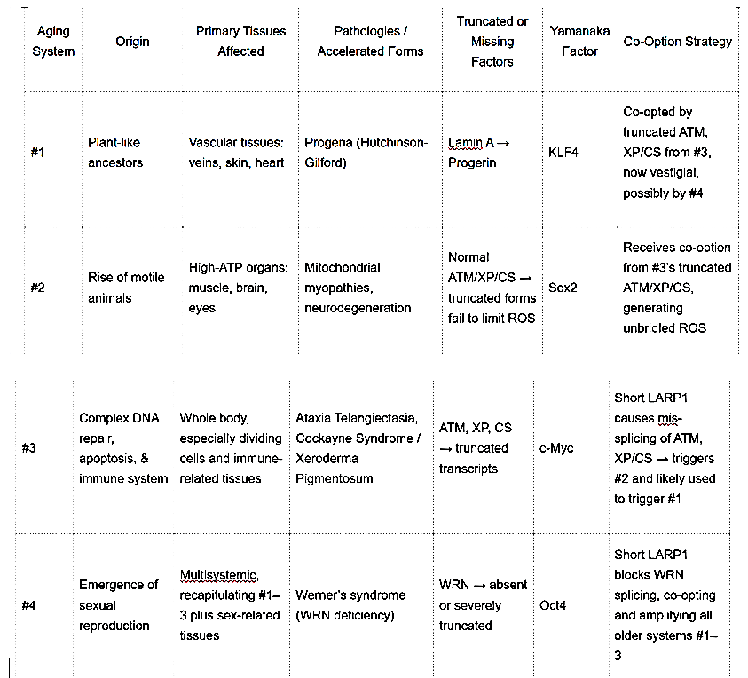
This table illustrates how each system builds on the last, with short LARP1 as a unifying factor.
Metabolic Imbalance and Epigenetic Regulation
The second post details the GABA–glutamate–αKG axis, where aging sees declining GABA, rising glutamate, and dropping αKG. GABA serum decline correlates with neural and metabolic dysfunction, diabetes, and cancer (possibly by inhibiting REST gene function) while glutamate excess fosters excitotoxicity. αKG, a TCA cycle intermediate, is crucial for DNA demethylation via TET and TDG enzymes. Its decline, by about 90%+ in serum with aging, impairs demethylation, leading to hypermethylation of promoters for many developmental and metabolic genes (36 of 48 aging genes Horvath’s first draft) , aligning with Horvath’s epigenetic clock data (Metabolic Imbalance and Aging GABA–Glutamate–αKG Axis).
The first post reinforces this, noting that Horvath’s 48 genes, including mRNA splicing factors and metabolic regulators, are hypermethylated with age. Metabolic shifts, like NAD⁺ and FAD depletion by CD38 and MAO-A & B, compound mitochondrial dysfunction, creating a feedback loop where metabolic decline triggers epigenetic dysregulation, further impairing metabolism. The additional post extends this by highlighting MAO-A and MAO-B’s role in depleting FAD, mirroring CD38’s NAD⁺ depletion, with both contributing to mitochondrial energy deficits (MAO-B Forces a Rethink of Darwin’s Legacy).
Initiating the Decline of GABA and αKG and the Role of Hormones
The four aging systems, coordinated by short LARP1, and exacerbated by death genes and hormonal changes, with a rise in LH, hCG, and possibly FSH leading to increased short LARP1 expression:
-
Short LARP1 and Mis-Splicing: Short LARP1 putatively mis-splices or suppresses mRNAs of critical proteins like WRN, ATM, and XP/CS, leading to their absence (WRN) or dysfunction. This dysfunction impairs differentiation in stem cells (WRN), DNA repair, ROS suppression, and structural integrity, setting off a cascade of aging-related pathologies that affect metabolic pathways, including maybe those for GABA and αKG production.
-
Death Genes and Cofactor Depletion: Enzymes like MAO-A, MAO-B, and CD38 increase with age, depleting FAD and NAD⁺, respectively. MAO-A and MAO-B can increase by 400–600% in the heart, and MAO-B by several hundred percent in the brain, leading to mitochondrial dysfunction. The depletion of NAD+ and FAD impairs the TCA cycle, reducing αKG production, and affects overall cellular energy, potentially impacting GABA synthesis (MAO-B Forces a Rethink of Darwin’s Legacy).
Notably, SP-1, one of Horvath’s aging genes, binds to the promoters of MAO-A and MAO-B, altering their expression with age. Additionally, SP-1 is involved in the transcriptional regulation of WRN, which is crucial for genome stability, and its expression is induced during puberty, linking the onset of sexual maturation (development) to the aging process.
-
Hormonal Changes Leading to Short LARP1 Expression: After age 50, hormones such as LH, FSH, and hCG increase significantly. FSH, which is more associated with general body aging (systems #1 and #2), and LH and hCG, which are more linked to reproductive and genetic stability/atrophy/cancer related aging (systems #3 and #4), both contribute to the aging process.
Specifically, FSH, which increases relatively more in men with aging, may predominantly trigger the aging symptoms related to systems #1 and #2, such as vascular and mitochondrial dysfunction, often seen as more “male-like” aging patterns. On the other hand, LH and hCG, which are more pronounced in women, may primarily activate systems #3 and #4, involving DNA repair, immune function, tissue atrophy, cancer, and reproductive aging, potentially more “female-like” aging patterns. How this occurs might be through altering the expression of short LARP1 in different tissue types?
These pro-aging hormones create a pro-inflammatory, possibly acidic, environment with increased free radicals, affecting epigenetic regulation and leading to increased expression of short LARP1. Short LARP1, in turn, mis-splices or suppresses mRNAs for key proteins across all aging systems, but the specific hormonal influences may bias its effects towards certain systems.
-
Impact on GABA and αKG: The decline in GABA might be due to reduced activity of the enzyme GAD (glutamic acid decarboxylase is an enzyme that catalyzes the decarboxylation of glutamate to gamma-aminobutyric acid) , possibly influenced by decreased pH levels influenced by increasingly acidic hormones LH, hCG, and/or FSH. Deceased serum GABA leads to increases in M1 macrophages and their production of CD38, depleting NAD⁺ further. The decline of αKG, also leads to shutting down about 36 of Horvath’s 48 genes, is linked to impaired TCA cycle function, exacerbated by FAD depletion from MAO-A and MAO-B. Hormonal acidity, potentially lowering intracellular pH, might alter enzyme activity, reducing GABA and αKG production and increasing glutamate. One intersting fact is tat the isoforms of LH that are produced after age 50 are relatively: larger, longer half lives, more bioactive, and much more acidic. It is not yet clear how or if acidic hormones are able to influence intracellular or mitochondrial pH.
-
M1 Macrophages and CD38: The decline of GABA and αKG contributes to a pro-inflammatory environment, promoting M1 macrophages, which express higher levels of CD38, depleting NAD⁺ and worsening mitochondrial function. This is supported by the user’s hypothesis that declining GABA and αKG lead to increased M1 macrophages or their CD38 production, possibly linking back to epigenetic changes via Horvath’s PAX5 gene protein, which represses CD38 and is downregulated with aging.
Interconnected Pathways and Feedback Loop
We propose a sequence:
- Aging, driven by the four evolutionary systems, leads to a rise in LH, FSH, and hCG as part of the natural aging/development process, particularly after 50, due to the decline of melatonin which suppresses LH , FSH and hCG and can act , at high doses , as effective birth control in human females at a dose of 75 mg/night. What causes the decline in melatonin? Possibly increased acidity in the pineal gland-this is still under study. It should also be noted that giving melatonin to menopausal women , even at doses as low as 6 mg a night had been found in some cases to reverse menopause and has even led to a pregnancy and birth several years after the cessation of menstruation (according to W. Pierpaoli).
- The increase in FSH primarily affects systems #1 and #2, while LH and hCG primarily affect systems #3 and #4, creating a pro-inflammatory, potentially acidic environment with increased free radicals, affecting epigenetic regulation and hypothetically leading to increased expression of short LARP1.Free radicals have been found to catalyze the demethylation of 5mC in normally regulated genes (non-TET/TDG controlled).
- Short LARP1 then may mis-splice or suppress mRNAs for WRN, ATM, XP/CS, and potentially Lamin A, activating all four aging systems, which further exacerbate the aging process. (Although the lamin A aging (loss of cellular differentiation) system appears to have become vestigial being replaced by some of Horvath’s 36 of 48 aging genes (transcription factors that maintain differentiation) that get turned off with aging .-see the 4 horsemen of aging blog post for more detail).
- Death gene proteins MAO-A, MAO-B, and CD38 increase, depleting FAD and NAD⁺, impairing mitochondrial function and reducing αKG production, while the hormonal environment reduces GABA production.
- The decline of GABA and αKG leads to epigenetic changes, with αKG decline hypomethylating, and thus shutting down, Horvath’s 36 of 48 genes controlled by TDG/TET enzymes, including PAX5, which suppresses CD38, increasing CD38 expression, and perpetuating the cycle.
This loop explains why aging accelerates: metabolic derangements cause epigenetic changes that worsen metabolism and activate death genes, with short LARP1 orchestrating the aging systems, initiated by the decline in melatonin and the rise in LH, FSH, and hCG, and modulated by SP-1’s regulation of MAO-A, MAO-B, and WRN.
Evidence and Support
- Horvath’s Genes: Table 1 from the first post ranks genes by association counts, with c-JUN (20 associations) and HDAC2 (12) as hubs, linking metabolism, epigenetics, and DNA repair, supporting interconnected pathways (Horvath’s Epigenetic Clock Analysis Across Biological Processes).
- Metabolic Data: The second post cites studies showing αKG supplementation reduces DNA methylation age by 8 years in humans (Metabolic Imbalance and Aging GABA–Glutamate–αKG Axis), linking metabolism to epigenetics.
- Evolutionary Layers: The third post’s table summarizes aging systems, with short LARP1’s role in co-option supported by LARP1’s presence in Horvath’s list and splicing database findings (Harvard’s spliceosome protein database derived from HeLa cells-where long LARP1 has no known spliceosome function (it was removed form the database after the curators were notified about it)) (Four Horsemen of Aging Evolutionary Systems).
- Death Genes: The additional post details MAO-A and MAO-B knockout phenotypes, with MAO-B knockout showing minimal early-life effects, supporting its status as a pure death gene, and therapeutic prospects like selegiline (which suppresses MAO-B at lower doses (5 mg in humans) and MAO-A and MAO-B at higher doses (40 mg/day in dogs) extending lifespan in animal models (MAO-B Forces a Rethink of Darwin’s Legacy).
- Hormonal and pH Hypotheses: The user’s information on hormonal changes and potential pH effects aligns with studies on aging and metabolic shifts, though direct evidence on pH changes needs further research.
Implications and Future Directions
This theory suggests aging is a coordinated program, not random damage, offering therapeutic targets:
- Restore αKG to enhance demethylation (Metabolic Imbalance and Aging GABA–Glutamate–αKG Axis).
- Inhibit CD38 and MAO-A/B to preserve NAD⁺ and FAD, and boosting NAD+ and FAD by vitamin B3 (niacin) and vitamin B2 (riboflavin) supplementation supporting mitochondrial function (MAO-B Forces a Rethink of Darwin’s Legacy).
- Target short LARP1 to prevent mis-splicing, potentially reversing multiple aging systems and using Yamanaka factors to reverse aging systems: (KLF4 for #1, Sox2 for #2, c-Myc for #3, Oct4 for #4).
- Supplement with melatonin to suppress LH and FSH and hCG (and melatonin also boosts progesterone), and supplement with DHEA and pregnenolone.
The complexity highlights the need for further research, especially on short LARP1’s regulation, MAO-A/B’s FAD depletion mechanisms, hormonal impacts on pH, and their evolutionary conservation, to validate this model and refine interventions.