About 6 years ago if you googled “The Streck Report” or “The Steck Report” you could quickly find a full copy of this classic 1937 study that showed the high doses Vitamin D are not dangerous.
I was looking for it recently and found that is has been scrubbed from the internet and hidden in two cases by expensive pay walls. So I am posting this study again for the benefit of all you Vitamin D3 researchers who are uncovering Big Pharma’s role in demonizing Vitamin D3.
AFTER POSTING THIS: WHEN I SEARCH STRECK OR STECK REPORT
THIS PAGE POPS UP ALMOST AS THE #1 RESULT ON page#1 ((OR CLOSE)
IN ALMOST ALL SEARCH ENGINES I TESTED: BING, YAHOO, DUCKDUCKGO, ETC
BUT WHEN I SEARCHED FOR IT IN GOOGLE
THERE WAS NOTHING FOR 6 RESULTS PAGES! WITH
JUST ONE LITTLE BLURB AT THE TOP OF GOOGLE’S PAGE 7 OF RESULTS: AS I SUSPECTED
GOOGLE IS CENSORED BY BIG PHARMA !
DO NOT USE GOOGLE WHEN SEARCHING FOR
ACCURATE HEALTH INFORMATION!
HERE IS THE STUDY AS FOLLOWS:
ANNALS OF
INTERNAL MEDICINE
VOLUME 10 JANUARY, 1937 NUMBER 7
FURTHER STUDIES ON INTOXICATION WITH
VITAMIN D * t
By I. E. STECK, M . D , H. D E U T S C H , A.B., C. I. R E E D , Ph.D., and H. C.
STRUCK, Ph.D., Chicago, Illinois
* Received for publication September 26, 1936.
From the Departments of Medicine and of Physiology, College of Medicine,
University of Illinois, Chicago.
This investigation at various stages has been supported in part by grants from Mead
Johnson and Company; the Graduate School Research Fund, University of Illinois; the
American Academy of Arts and Sciences; the Wisconsin Alumni Research Foundation;
the Committee on Therapeutics of the American Medical Association; and the Phi Rho
Sigma Medical Fraternity.
W I T H the extensive application of massive doses of vitamin D as a
therapeutic agent in various clinical conditions numerous
criticisms have arisen which may be summarized as follows:
“1. Hypervitaminosis D may produce symptoms of hyperparathyroidism.
2. The therapeutic use of vitamin D is rational only in conditions of
known deficiency.
3. Animals experimentally treated with vitamin D concentrates have
shown extensive calcium deposits in various tissues, and other pathological
changes have been found. There is, thus, danger of permanent injury to
human subjects.”
It is not the purpose of this paper to discuss the therapeutic value of concentrated vitamin D in any clinical condition. Only by more extensive clinical investigation can its therapeutic value be established finally.
That vitamin D in massive doses may be toxic to any individual, animal or human, has been recognized in all stages of this series of investigations which was begun early in 1929, and its administration to human subjects has been governed accordingly. It is the purpose of this paper to presentevidence bearing on these questions.
The first of these questions has been discussed very thoroughly by Shelling, who concluded that the preponderance of evidence is against the view that the activity of vitamin D is dependent upon the functional integrity of the parathyroids and in favor of the existence of an antagonism between the two. The second criticism is purely speculative. If this idea held true very few therapeutic procedures now in use could be justified.
In considering the nature of the action of massive doses of vitamin D itis assumed that the action is very different from the physiological effects of small doses. It is so different, in fact, that the concentrated material may be considered tentatively as a different substance. At the same time there is some legitimate doubt as to the justification for this attitude.

Early experience with impure preparations of vitamin D, particularly abroad, has led to a great deal of misunderstanding and fear of overdosage on the part of those who have had little acquaintance with the fundamental mechanisms involved. This point has been adequately discussed by Bills. Suffice it to say that most of this earlier work must be disregarded when considering the effects produced by the highly purified preparations now available.
954 E. STECK, H . DEUTSCH, C. I. REED AND H . C. STRUCK
E X P E R I M E N T S ON TOXICITY OF V I T A M I N D FOR DOGS
The effects of massive doses of vitamin D must be judged on the basis
of the dose per unit body weight and not on the absolute size of the dose.
When considered in this light the order of increasing susceptibility appears
to run as follows: rat, dog, human, rabbit, with little difference between the
dog and human, while the rat is very much more resistant, and the rabbit
much less so.
In the experiments on dogs, vitamin D was administered in the form of a
solution of activated ergosterol (Vitamin D2) in corn o i l (1,000,000 units per gm.)
Or of calciferol dissolved in corn oil.
Most of the administration was done
orally. A few animals received intravenous injections in which form the
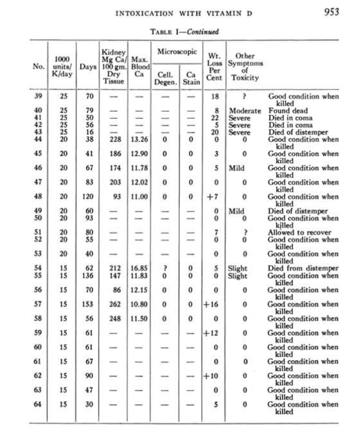
material is slightly more effective. The results of experiments on 64 adult
healthy dogs are shown in table 1. The daily dose ranged from 15,000 to
500,000 units per kilogram of body weight as shown in the first column.
An effort was made to adjust the dose to decreasing body weight so that the
ratio between the dose and the weight of metabolizing tissue remained fairly
constant.
The figures in the second column represent the actual number of days of
administration of the vitamin D and, therefore, the number of days the
animals survived the treatment. Usually those that did not die were killed
on the day following the last dose, others within two to three days. With
amounts greater than 50,000 units daily the average survival period was 12
days; with amounts between 20,000 and 50,000, 39 days; and with 20,000
units or less, 68 days.
At death, tissues were taken for chemical analysis and for microscopic
examination. In an earlier report (10) on analyses of 13 different tissues in a
series of dogs, it was shown that the kidney is the most vulnerable of any
tissue to the calcifying action of vitamin D. In order to conserve space,
only the figures for the calcium content of the kidney are included. In all
other tissues the findings were very inconstant. Analyses of tissues of nor-
mal dogs have shown a calcium content in the kidneys ranging from 29 to
301 mg. per 100 gm. of dried tissue with a mean of 85 mg. The calcium
content in the kidneys of 33 of the 64 dogs is shown in the third column
of table 1.
With a daily dose above 50,000 units per kg. the average content of calcium
in the kidney was 564 mg. per 100 gm. of dried tissue; between 20,000
and 50,000, 921 mg.; and with 20,000 units or less, 183 mg. The lower
average in the first group may possibly be related to the shorter period of
survival. The average in the third group is obviously lower because the
dose was not great enough to cause as much deposition.
In the fourth column the figures represent the maximum concentration of
9 5 6 I. E. STECK, H . DEUTSCH, C. I. REED AND H. C. STRUCK
cellular injury nor of excessive calcium deposition in any other tissue
examined. From evidence obtained on 12 other animals not included in the table,
It appears that considerable loss of weight may occur before there is any
evidence of injury to cells, but this was not invariably the case.
Our observations do not confirm those of Appelrot to the effect that
hypervitaminosis D produces medial thickening in arteries. In only four of
the 64 dogs examined, i.e., numbers 18, 21, 27 and 29, was this observed.
In each case there was some medial thickening in the aorta and occasionally
in smaller arteries.
Loss of weight was marked in 36 out of the 43 dogs receiving more than
20,000 units per kg. per day. One retained constant weight, one gained 17
per cent. As previously suggested, the early weight loss appears to be due
mainly to impoverishment of fat depots. When dog No. 28 was examined
at autopsy it was almost impossible to find any macroscopic fat deposits.
This dog lost 48 per cent of the original weight, yet showed only mild
objective symptoms of toxicity and was lively and active at the termination
of the experiment. There was practically no microscopic evidence of cell
injury. The plasma calcium was maintained at a higher average level over
the period of the experiment than in any other animal in our experience.
On the other hand, dog No. 21 lost only IS per cent of the initial weight,
although the kidney content of calcium was higher than in any other animal.
These two animals serve to show clearly that weight loss is not an inevitable
accompaniment of fatal intoxication in dogs.
One of the characteristic features of fatal hypervitaminosis D is the
premortal coma. This condition is usually, though not always, preceded by
partial paralysis, slow, shallow respiration, fine, thready, rapid pulse, saliva-
tion, and often by psychic changes of such a nature that a previously tame,
friendly dog may become unmanageable and even vicious. Very often the
symptoms resemble those following an injection of oil of wormwood. This
condition may persist for several days but usually appears from two to three
hours before death. It is probable that all of the dogs labelled ” found
dead ” passed through this stage during the night, but the actual train of
symptoms could not be observed. None of the animals that were killed were
in this stage.
The objective symptoms of toxicity were much the same as those previously described for the human, such as weakness and lassitude, anorexia,
polydipsia, polyuria, psychic disturbances, diarrhea. Bloody feces were
passed by 11 of the 64 dogs. In addition, petechial hemorrhages were found
in the mucosa of the stomach and intestines at autopsy.
With eight exceptions all of the 43 dogs receiving more than 20,000
units per kg. per day died spontaneously. Nine of these died from dis-
temper. Of the nine, three were in such a condition that early death in coma
was predictable. Of the eight exceptions, one was allowed to recover. Of
INTOXICATION WITH VITAMIN D 957
the seven that were killed, two would probably have died. The other five
would probably have recovered with cessation of the treatment.
Among the 20 dogs receiving 20,000 units or less per kg. there were no
evidences of cell injury, insignificant weight loss, very little evidence of toxic
symptoms, and with the exception of two dogs that died from distemper and
one that was allowed to recover all were in good condition when killed.
Thus, it may be concluded that vitamin D up to 20,000 units per kg. per
day for periods ranging up to 153 days is not seriously injurious to normal
dogs. In greater amounts there is a wide range of susceptibility entirely
unpredictable from any data at present available.
That the toxic eflfects may be characterized as true hypervitaminosis D
is proved by the fact that in four instances (numbers 7, 17, 29, 34) the
vitamin preparation administered was crystalline calciferol (40,000,000 units
per gm.) in solution in corn oil. This preparation contained no toxic by-
products and yet the results were quite comparable with those from the same
dose of activated ergosterol which may contain some inert material but
practically no toxisterol.
In another series of experiments the dogs were brought to a stage of
extreme toxicity with vitamin D and the administration was then discontinued.
The state of toxicity was manifested by loss of weight, anorexia,
listlessness and paralysis, and, in six animals, prostration. After varying
intervals, when there was objective evidence of complete recovery, the ani-
mals were killed and subjected to the same examination as in the other series.
The results are shown in table 2. In the first column are shown the periods
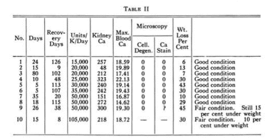
f administration of the vitamin, in the second the daily dose per kg., and in
the third the recovery interval allowed. Actually in the dogs No. 4, 6,
7 and 8 complete recovery of weight was accomplished in 53, 86, 73 and 60
days respectively. Animals No. 9 and 10 were killed before weigh
9 5 8 I. E. STECK, H. DEUTSCH, C. I. REED AND H. C. STRUCK
recovery was complete because both were definitely overweight when the
experiments were begun.
Eight additional dogs were originally included in this series but all of
these were in the terminal stages of toxicity described above when the treat-
ment was discontinued, and all died within two to seven days. Six of these
were already prostrate when administration was discontinued.
In all but one (dog No. 4) the kidney content of calcium was within
the normal range, although all showed figures in the higher limits of the
normal range except Nos. 2 and 7. The average weight loss approximated that
in comparable dosage ranges in the first series.
The maximum concentrations of plasma calcium observed were all high.
Microscopic examination of the tissues showed no definite evidence of calcium
deposition and no cellular injury. In Nos. 2 and 5 there was some
distortion of the collecting tubules in the kidney by fibrosis. No vascular
lesions were found.
From these experiments it appears that dogs may recover from extreme
stages of toxicity and that whatever tissue injury occurs may be reparable.
In extremes the result may be fatal before repair can be effected.
In the experiments on animals reported by others, so far as the potency
of the dose can be calculated, toxicity has occurred only with doses above
20,000 units per kg. per day. Since the relation of dosage to weight has not
been stressed before, many other workers have failed to record data which
would make it possible to calculate the dose in terms of units per kilogram.
In both of our series it is apparent that the total amount of vitamin ad-
ministered is not the most important determinant of the degree of toxicity
since large doses have been tolerated well over long periods while smaller
doses have produced drastic effects in a few days. Nor is the magnitude of
the daily dose the sole factor. In general, both dogs and human subjects
appeared to be less sensitive during winter months
INTOXICATION WITH VITAMIN D 959
It has been emphasized many times that the diet is a very important
factor in conditioning toxicity. All of these animals were kept on the stock
kennel diet throughout and no variations were introduced at any time.
It is also apparent that the concentration of plasma calcium is not closely
correlated with toxicity. Many investigators have been inclined to use the
terms ” hypercalcemia ” and ” toxicity ” interchangeably. Many experiments
have been done which clearly disprove a causal relation between these
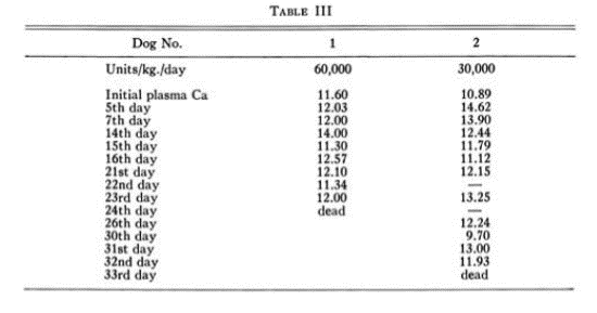
conditions but the two experiments reported in table 3 will serve to illustrate
the point. Both of the animals showed some hypercalcemia, but it was by no
means pronounced. Number 1 received twice the dose of number 2, but
except for the earlier death there was no striking difference. When one
contrasts these figures with the high figures for some of the animals that
survived as in tables 1 and 2, it is a fair assumption that hypercalcemia per se
is not the cause of toxicity.
OBSERVATIONS ON H U M A N S U B J E C T S
The enormous absolute doses of vitamin D that have been administered
to human subjects have naturally aroused some question. If, however, one
bears in mind that human and canine susceptibility seem closely approxi-
mated, and if one gives due consideration to the weight/dosage relationship,
it appears that few of the human subjects have ever received amounts com-
parable to the highest doses tolerated by the dogs of this series.
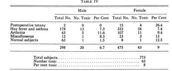
Table 4 shows the status of 773 human subjects who have received
amounts of vitamin D above 100,000 units daily. The doses routinely given
ranged upward from 200,000 units total daily dose for periods ranging from
seven days to five years. With such a dose a 50 kilo subject would receive
only 4,000 units per kg. per day. With 300,000 units a 60 kg. subject would
receive 5,000 units per kg. per day. This table, of course, does not take into
account the varying dosages, so that one cannot draw conclusions as to comparative susceptibility in the various groups of subjects. However, it appears
9 6 0 I. E. STECK, H. DEUTSCH, C. I. REED AND H. C. STRUCK
from other statistics too voluminous to include here, that the order
of decreasing susceptibility among the different groups of patients is:
arthritis, normal subjects, hay fever alone, hay fever with asthma, tetany.
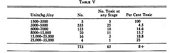
In table 5 the incidence of toxicity at each range of dosage is shown.
This analysis does not take into account the duration of administration since,
as was indicated by the experiments on dogs, the total amount does not
seem to be the most important factor. Nor does it take into account the
effect of simultaneous administration of yeast as a protective measure which
was done with many human subjects. This procedure has already been
discussed rather fully.4’5 The first five subjects may be disregarded since
they became nauseated from corn oil as readily as from viosterol, so that
they do not represent true hypervitaminosis D, but rather some kind of sen-
sitivity not related to the vitamin. It is probable that some of those sub-
jects included in the toxic groups at higher dosage were of this type. The
shortest period of administration that produced intoxication in the group on
3,000 to 5,000 units per kg. per day, was 87 days. Since the condition in
this instance developed very suddenly and without any weight loss, it is
possible that some unrecognized disturbance rendered the subject tem-
porarily more susceptible. On discontinuance of administration of vitamin
D prompt recovery occurred, and four days later the treatment was resumed
and continued four months without further disturbance.
In the group on 6,000 to 7,000 units per kg. per day the shortest period
for development of toxicity was 60 days. In this instance there was a loss
of four pounds in two days. The condition was abated sufficiently to re-
sume only after eight days without treatment. A second mild intoxication
occurred three months later.
In the last two groups the high doses were not continued beyond 10 days
except in one case, regardless of whether intoxication occurred. In this
case one of the authors, a normal subject, took 35,000 units per kg. per day
(3,000,000 units total daily) for 15 days without any evidence of dis-
turbance of any kind.
Vrtiak and L a n g 7 have recently reported 100 per cent incidence of tox-
icity in a series of 22 human subjects to whom massive doses of Vitamin D
INTOXICATION WITH VITAMIN D 961
were administered. The discrepancy between this high incidence and the
relatively low incidence in our series is difficult to explain at present.
The symptoms of hypervitaminosis D in human subjects were described
fully in an earlier paper.4 It remains now to correlate these findings with
the experimental results on animals. Tentatively we suggest that the course
of events is as follows: First, cellular degeneration occurs, more commonly
in the kidney. Concurrently there may be loss of weight and other objective
symptoms. If weight loss occurs before other symptoms it is probably due
solely or mainly to increased fat catabolism. Second, deposition of calcium
occurs at the sites of cell injury; apparently this does not occur except as a
secondary result of such injury. Third, up to advanced stages of toxicity
these processes may be reparable in dogs if the vitamin D administration is
discontinued.
From the results of previously reported work it appears that the
calcium removed from the tissues during recovery is excreted in the urine.
At least, repair is not complete until the urine calcium excretion becomes
normal.
The increased excretion of calcium that usually takes place under
massive administration of vitamin D in both human subjects and dogs is not,
however, due solely to removal of the microscopic deposits in the soft tissues,
because the increase in the urine begins before there is any microscopic or
chemical evidence of excessive deposition in soft tissues. The source of this
initial increase in the urine has not yet been determined, but from Shelling’s
discussion it probably comes from the trabeculae of the bones. Generally
the average level of blood calcium is decreased after the excretion is in-
creased.
We have made no examination of blood pressure in dogs, but a large
number of the human subjects were examined at frequent intervals over long
periods. Since there have been no significant changes the data are omitted.
In most instances there seems to be some tendency to a slight decrease in the
general level. This, of course, does not eliminate the possibility of medial
proliferation in the arterioles.
Our human subjects ranged in age from 17 to 76 years. Older subjects
were generally less readily intoxicated but recovered less readily when in-
toxication did occur, and seemed to be somewhat more sensitive thereafter.
There are on record only two instances of death in human subjects,
certainly due to hypervitaminosis D, since the more highly purified preparations became available.
One of these, recently reported by Thatcher, was
probably a case of idiosyncrasy to vitamin D. It is difficult to determine
from the report the unit dosage. However, it is clear that the administration was continued after intoxication was markedly developed. We wish to stress that administration of vitamin D should be discontinued at once when the symptoms of intoxication appear. Neither animal
nor human subjects in our experience have ever recovered from the toxic
condition while administration continued.
9 6 2 I. E. STECK, H. DEUTSCH, C. I. REED AND H. C. STRUCK
The other case has not yet been reported in detail in the literature, but
the reports of the coroner and the attending physician 15 reveal the following
facts. A physician, aged 74, weight 290 pounds, undertook self-medication
with a concentrated solution of activated ergosterol. Owing to an error in
calculation of the dosage he received 2,300,000 units daily for 18 days or
approximately 18,000 units per kg. per day, a dose 10 times that intended.
Since he was very obese the dose per kilogram of actively metabolizing tissue
was much greater. The symptoms described were quite typical of hyper-
vitaminosis D, with hypercalcemia, so that there is no doubt that in this case
the treatment was the immediate cause of death. However, the presence
of generalized arteriosclerosis suggests that this was a fundamental handicap
to his recovery after discontinuing the treatment.
The administration of similar or larger amounts in our series without
serious disturbance should not be interpreted to mean that such treatment
can be undertaken without caution. In fact, our experience indicates clearly
that administration of massive doses of vitamin D should not be undertaken
for any cause except under the careful supervision of a physician who can
and will carefully check the patient’s condition at frequent intervals and who
will see to it that the treatment is discontinued promptly on the appearance
of the first signs suggestive of toxicity.
It is probable that any suggestion of kidney dysfunction should constitute an absolute
contraindication. Until further information is available
arteriosclerosis also should probably be considered a contraindication. Cosequently,
this form of treatment should be administered to older subjects only with extreme caution.
Nevertheless, if these precautions are observed, massive doses of vitamin
D may be utilized therapeutically as safely as many other agents administered daily.
That its misuse has resulted in death should not prejudice its
controlled use under circumstances of possible value.
In view of the extensive experience in administration of vitamin D to
human subjects with a relatively low incidence of toxicity, and the correlation of the
results of animal experiments with the observations on human
subjects, we believe that the burden of proof now rests on those who main-
tain the undesirability of the use of this form of therapy. Its actual practical value
in particular clinical conditions will, of course, be determined only
by more extensive clinical experience.
It must be admitted that the mechanism of toxicity is still unexplained.
Our findings do not agree entirely with those of Ham and Lewis.16 It is
possible that the dose of approximately 600,000 per kg. per day or more,
which these observers administered to rats, was a factor in producing a different
type of lesion. Up to the present time conceptions of the physiology
of vitamin D have perhaps been too circumscribed because of its striking
effect on calcium metabolism. That the thyroid plays an important part in
the action of vitamin D is indicated by the results of another investigation.
INTOXICATION WITH VITAMIN D 963
Also, it appears that the pathologic effects are greatly accentuated in a hypo-
thyroid state. This may be an important factor in the variability in sensitivity to
intoxication.
The results obtained by Gelfan on isolated frog muscle suggest that
vitamin D exercises a catalytic effect in peripheral tissues. If this should
be confirmed, it might serve as a forward step in explaining the injury to
isolated cells in peripheral tissues. It is conceivable that the metabolism of
individual cells might be accentuated to such a point as to result in the dis-
integration of the cells themselves. We have not been able to recognize the
cellular changes preliminary to disintegration so that we can offer no ex-
planation of the actual nature of this process.
SUMMARY AND CONCLUSIONS
1. Observations on 64 dogs and 773 human subjects receiving massive
doses of vitamin D have been made and data recorded as to dose per unit of
body weight, and on the nature of the process of intoxication.
2. Both human subjects and dogs generally survive the administration of
20,000 units per kilogram per day for indefinite periods without intoxication.
3. Hypervitaminosis D first produces cell injury followed by calcium
deposition. This process is reversible and reparable if administration is
discontinued promptly.
4. Intoxication for short periods does not result in any permanent injury
that can be recognized by the methods employed in this investigation.
ADDENDUM. After this manuscript was written there came to our attention a very
comprehensive study by Cowdry and Scott1 9 on normal monkeys under treatment with
vitamin D in which it was suggested that pathological changes may occur in tissues without
clinical symptoms. However, it was also suggested that there might be a species peculiarity.
BIBLIOGRAPHY
1. REED, C. I., and SEED, L.: The treatment of clinical tetany with irradiated ergosterol,
Endocrinology, 1933, xvii, 136-148.
2. RAPPAPORT, B. Z., and REED, C. I.: Viosterol of high potency in seasonal hay fever and
allergy, Jr. Am. Med. Assoc, 1933, ci, 105-109.
3. RAPPAPORT, B. Z., HATHAWAY, M. L., REED, C. I., and STRUCK, H. C : The treatment of
hay fever and asthma with viosterol of high potency, Jr. Allergy, 1934, v, 541-553.
4. REED, C. I.: The symptoms of viosterol overdosage in human subjects, Jr. Am. Med.
Assoc, 1934, cii, 1745-1748.
5. DREYER, I , and REED, C. I.: The treatment of arthritis with massive doses of vitamin D,
Arch. Phys. Therapy, 1935, xvi, 537-540.
6. CRIMM, P. D , et al.: Vitamin therapy in pulmonary tuberculosis, Am. Rev. Tuberc,
1932, xxvi, 112-123; Ibid., 1933, xxviii, 202-216; Am. Jr. Med. Sci, 1934, clxxxvii,
557-562; Jr. Lab. and Clin. Med., 1934, xix, 966-971.
7. VRTIAK, E. G., and LANG, R. S.: Observations on the treatment of chronic arthritis with
vitamin D, Jr. Am. Med. Assoc, 1936, cvi, 1162-1163.
8. SHELLING, D. H.: The parathyroids in health and disease, 1935, C. V. Mosby Co., St.
Louis, Mo.
9. BILLS, C. E.: Physiology of the sterols, including vitamin C, Physiol. Rev., 1935, xv,
1-97
9 6 4 I. E. STECK, H . DEUTSCH, C. I. REED AND H . C. STRUCK
10. REED, C. I., D I L L M A N , L. M., THACKER, E. A., and K L E I N , R. I . : The calcification of
tissues by excessive doses of irradiated ergosterol, Jr. Nutr., 1933, vi, 371-381.
11. APPELROT, S.: Hypervitaminosis D and blood pressure in dogs, Am. Jr. Physiol., 1933,
cv, 294.
12. REED, C. I., THACKER, E. A., D I L L M A N , L. M., and W E L C H , J. W . : The effects of’
irradiated ergosterol on the metabolism of normal dogs, Jr. Nutr., 1933, vi, 355-370.
13. H A T H A W A Y , M. L., RAPPAPORT, B. Z., REED, C. I., and STRUCK, H. C.: A study of blood
constituents in pollenosis with and without treatment, Jr. Allergy, 1936, viii, 1-21.
14. THATCHER, L . : Hypervitaminosis D, Lancet, 1936, i, 20-22.
15. KERR, W. J., and CARR, J. L . : Personal communications.
16. H A M , A. W., and L E W I S , M. D . : Experimental intimal sclerosis of the coronary arteries
of rats, Arch. Path., 1934, xvii, 356-361.
17. D E U T S C H , H., REED, C. L, and STRUCK, H. C.: The role of the thyroid in the calorigenic
action of vitamin D, Am. Jr. Physiol., 1936, cxvii, 1.
18. GELFAN, S.: The effect of viosterol upon oxygen consumption of frog’s muscle, Am.
Jr. Physiol, 1935, cxiii, 464-466.
19. COWDRY, E. V., and SCOTT, G. H . : Effect on monkeys of small doses of a concentrated
preparation of viosterol, Arch. Path., 1936, xxii, 1-23